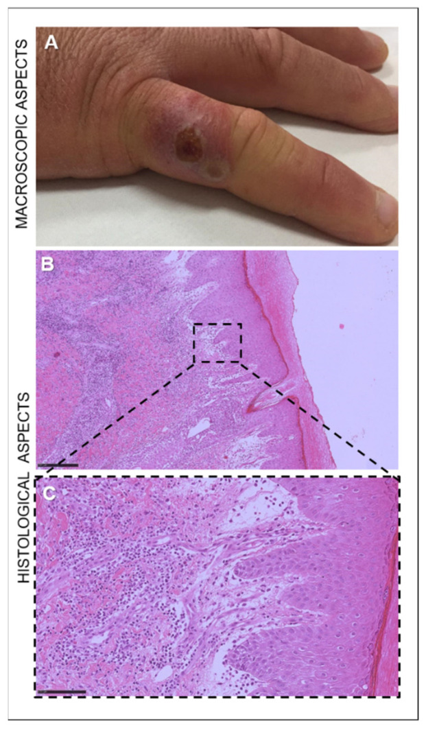
Figure 3.
Nodule on the right little finger of a 30-year-old sheep farmer infected with ORFV. (A) Targetoid nodule with a necrotic center, a white ring, and peripheral erythema. (B) Microscopical appearance of the skin of the ORF-infected man, characterized by marked acanthosis and hyperkeratosis. Hematoxylin and eosin staining (H&E). Original magnification 50×. Scale bar = 250 µm. (C) In the dermis, vascular proliferation with granulocytic and eosinophilic infiltration. Hematoxylin and eosin staining (H&E). Original magnification 400×. Scale bar = 100 µm.

