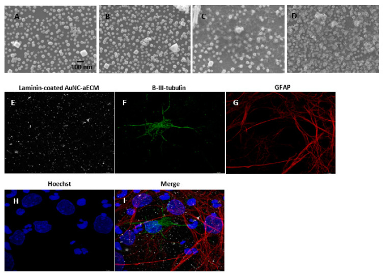
Figure 1.
Characterization of AuNC-aECMs and laminin-coated AuNC-aECMs. (A–D) Scanning electron microscopy of AuNC-aECMs. The quantity of AuNCs can be controlled by reaction buffer solution and the concentration of AuNC colloidal solution (A: 12.8 fmol, 32 AuNCs/μm2 and B: 25.6 fmol, 54 AuNCs/μm2). The AuNC-aECMs were coated with laminin (C: 32 AuNCs/μm2 and D: 54 AuNCs/μm2). The cells growing on laminin-coated AuNC-aECMs (32 AuNCs/μm2) were analyzed with immunostaining assay and fluorescent images were obtained using Leica—Lightning Light Laser Confocal Microscope SP8X (E–I). (E) Laminin-coated AuNC-aECM, (F) β-III-tubulin immunostaining (neuron marker), (G) Glial fibrillary acidic protein (GFAP) immunostaining (astrocyte marker), (H) Hoechst staining, (I) Merged image.

