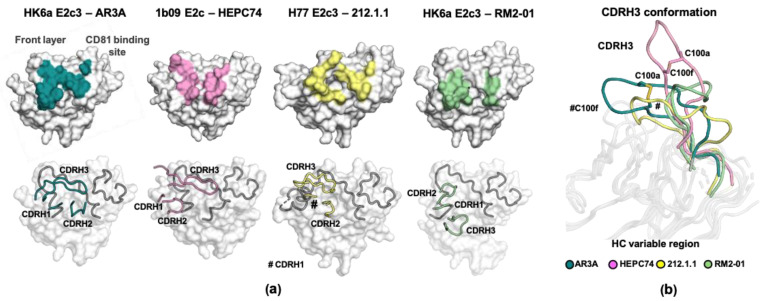
Figure 3.
The binding mode of AR3-targeted VH1-69-encoded bnAbs. (a) Structural comparison of the epitopes and the CDR positions of the bnAb subgroups. AR3A is representative of the AR3A/B/C/D, U1 and HC11 group; HEPC74 of the HEPC74 and HEPC3 group; 212.1.1 of the 212.1.1 and HC1AM group; and RM2-1 of the RM2-1 and RM11-43 group. Top: the epitopes of AR3-targeted VH1-69-encoded bnAbs. E2c structures are shown in surface representation and antibody footprints are colored and labelled. Bottom: the position of the HC CDRs in the E2c–Fab structures. E2c domains are shown in surface representation with the front layer and CD81 binding loop as an illustration and colored in gray. The HC CDRs are also illustrated. (b) Conformation of CDRH3 of AR3A, HEPC74, 212.1.1, and RM2-01 from the E2–bnAbs complexes. The intra disulfide bond in the CDRH3 of AR3A and HEPC74 is shown in stick representation.

