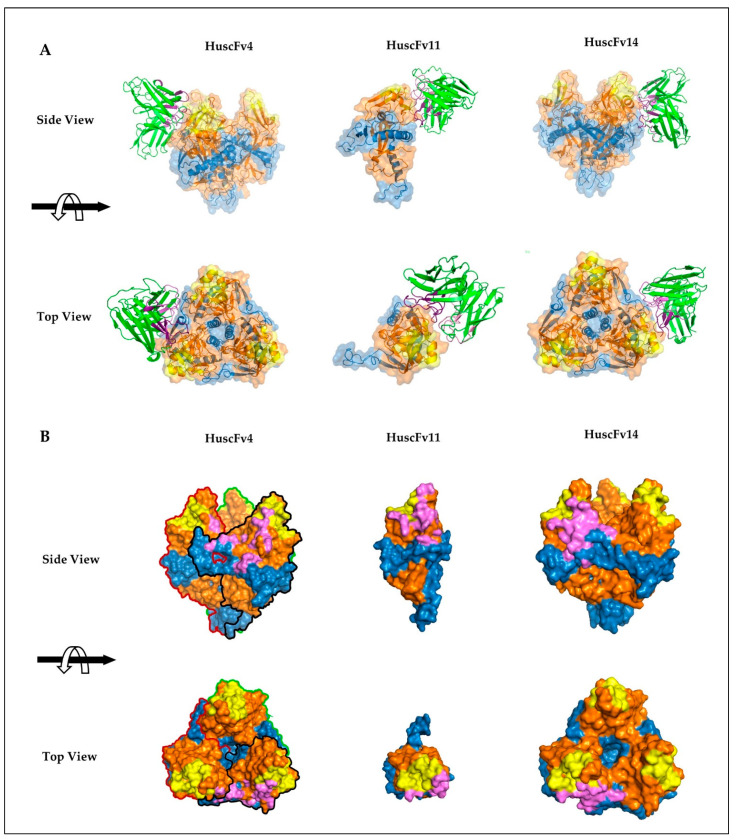
Figure 5.
Presumptive binding of HuscFvs to GPcl predicted by computerized homology modeling and intermolecular docking. (A) Interactions of HuscFv4 and HuscFv14 to trimeric GPcl (PDB: 5F1B) (left and right panels, respectively) and HuscFv11 to monomeric GPcl (PDB: 5HJ3) (middle panel). The HuscFv-GPcl complexes are shown as the side view (upper panel) or top view (lower panel). The HuscFvs are colored in green, in which CDRH and CDRL loops are colored as dark and light purple, respectively. GP1, GP2, and RBS of the GPcl are in orange, blue, and yellow, respectively. (B) Footprints in HuscFv-GPcl complexes as described in (A). Contact surface areas between the HuscFvs and the GPcl are colored in purple. The GP1, GP2, and RBS of each GPcl protomer are colored as described in (A). Solid black, red, and green lines indicate boundaries of individual GPcl protomers.

