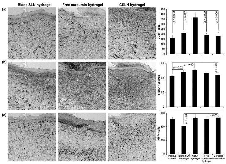
Figure 11.
Effect of CSLNs on the angiogenesis (CD31), remodeling (α-SMA), and proliferation (Ki67) in skin wound after healing at day 11. Representative images of blank SLN hydrogel, free curcumin hydrogel, and CSLN hydrogel are shown for different staining’s (top to bottom and left to right, respectively). Serial sections were stained separately with anti-CD31 (a), anti-α-SMA (b), and anti-Ki67 (c), and visualized with a 10X objective. The number of +Ve cells/area obtained by ImageJ analysis were compared between all the groups (right panel, top to bottom). Data are represented as mean ± SEM. Statistical significance was determined by ANOVA followed by Tukey HSD. Arrows indicate +Ve cells for respective staining.

