Figure 9.
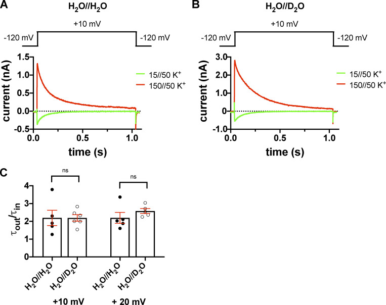
Inward and outward K+ currents in H2O- or D2O-based extracellular solutions in the T449A Shaker-IR mutant. (A and B) Macroscopic currents were measured in voltage-clamped inside-out patches excised from tsA201 cells and normalized to their respective peak currents. The extracellular (pipette-filling) solution contained 50 mM K+ in H2O (A) or D2O (B), and the intracellular (bath) solution contained 150 mM K+ (red traces) or 15 mM K+ (green traces). The holding potential was −120 mV; 1.0-s-long depolarizing pulses to +10 mV were applied to activate the channels and record inward K+ current (green curves) or outward K+ current (red curves). The voltage protocols are shown above the corresponding raw current traces. (C) Inactivation time constants of the currents were determined by fitting a single-exponential function to the decaying part of the currents. Bars and error bars indicate the mean ± SEM of the ratio of the inactivation time constants measured for the outward and inward currents (τout/τin) at +10 mV and +20 mV test potentials in H2O//H2O solution (filled circles) and extracellular D2O (H2O//D2O, empty circles). Symbols indicate the individual data points (n = 4–6).

