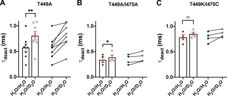
Figure S2.
Effect of extracellular D2O substitution on the deactivation kinetics of Shaker-IR channels. (A–C) Deactivation kinetics of T449A (A), T449A/I470A (B), and T449K/I470C (C) Shaker-IR channels, expressed in tsA201 cells, was analyzed in the presence (H2O//D2O) and absence (H2O//H2O) of extracelular D2O. The cells were depolarized to +50 mV from a holding potential of −120 mV for 15 ms in outside-out configuration. Tail currents (Itail) were evoked by stepping the test potential back to −120 mV at the end of the 15-ms-long depolarization (see Fig. 3). The deactivation time constant (τdeact) was determined by fitting a single-exponential function to the decaying tail currents: where A is the tail current amplitude (pA), t is time (ms), τdeact is the deactivation time constant (ms), and C is the y offset. The deactivation time constant for a particular cell was defined as the average of time constants obtained for at least three repolarizing pulses repeated at every 15 s in a sequence. Bars and error bars indicate mean ± SEM (n ≥ 4) of τdeact for the indicated clones in H2O//H2O (filled symbols) and H2O//D2O (empty symbols). Symbols indicate individual data points (circles, T449A; down triangles, T449A/I470A; up triangles, T449K/I470C). Paired data are shown on the right part of each panel. Asterisks indicate significant differences (*, P < 0.05; **, P < 0.01).

