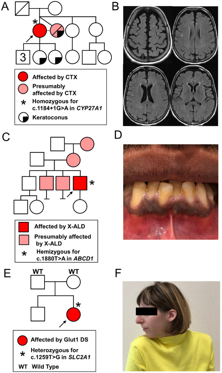
Figure 1.
Family trees and phenotypic data of the three patients: (A,B) proband 1: family pedigree (left panel) and axial views of brain MRI, FLAIR images showing non-confluent bilateral periventricular and semi-oval center white matter hyperintensities (right panel); (C,D) proband 2: family pedigree (left panel) and a picture of the mouth showing gingival hyperpigmentation (right panel); (E,F) proband 3: family pedigree (left panel) and a photograph showing cervical dystonia with right torticollis, as well as microcephaly. CTX, cerebrotendinous xanthomatosis; Glut1 DS, glucose transporter type 1 deficiency syndrome; X-ALD, X-linked adrenoleukodystrophy.

