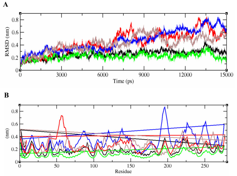
Figure 5.
RMSD and RMSF plots generated for the epitope-HLA complexes of Wuhan, England, USA, Indian, and South African variants. (A) represents the unstable RMSD values of the complex from England, India, South Africa, USA, and the Wuhan isolates in green, black, brown, red, and blue respectively.The epitope/HLA combinations of England and Indian strains were found to be more stable than that of others. (B) represents the fluctuation patterns of the protein–peptide complexes of all five SARS-CoV-2 variants analyzed with their RMSF values given in nm. The amino acid residues of Wuhan strain displayed a maximum deviation in the fluctuation map up to 0.8 nm.

