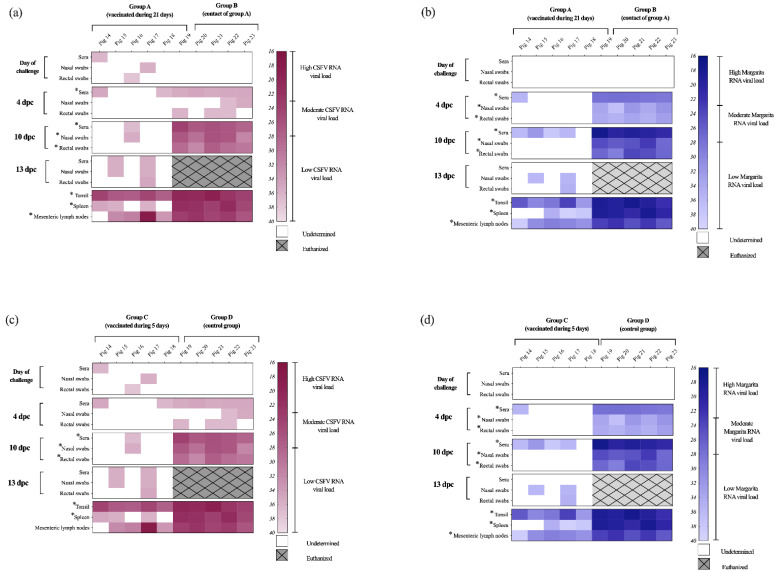
Figure 6.
CSFV RNA detection at different time points after challenge in samples and tissues. The samples were analyzed by RT-qPCR for (a,c) the CSFV [24] and (b,d) Margarita strain RNA [20] viral load at different time points during the 13 dpc (or until euthanasia). White area indicates negative samples. Euthanized animals are represented in grey square with hatches. Asterisk symbol (*) indicates statistically significant differences of the Ct values between the groups (p < 0.05).

