. 2020 Jan 10;11(7):1750–1760. doi: 10.1039/c9sc05623h

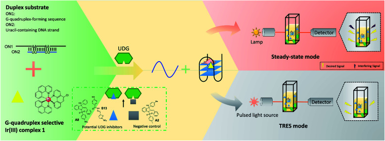
Fig. 1. Schematic diagram of the novel UDG inhibitor screening method. In the assay mechanism, the G-quadruplex-forming motif (ON1, 5′-G3TAG3A3T2CT2A2GTGCG3T2G3-3′) is hybridized initially with a partly complementary, uracil-containing DNA sequence (ON2, 5′-CGCACU2A2GA2U2TC-3′) to form a double-stranded DNA substrate. The addition of UDG excises uracil bases from ON1–ON2, forming abasic sites on ON2 which releases ON1. ON1 converts into a G-quadruplex motif that is then bound by the iridium(iii) complex 1 with enhanced emission. In the presence of a UDG inhibitor, less ON1 would be liberated, resulting in a weaker luminescence intensity from probe 1. However, this decrease might become completely swamped by the background fluorescence, resulting in a false negative result (upper panel). By using TRES, the short-lived fluorescence of inhibitors would be eliminated, allowing the reducing in luminescence intensity of probe 1 to be clearly detected (lower panel).

Fig. 1