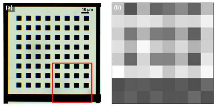
Figure 8.
(a) Optical image of the EBL pattern of 6.4 µm squares regular spaced at 6.4 µm, with the observed region highlighted in red. (b) Image reconstructed with the microscope prototype based on the 8 × 8 5 µm LED array chip (Led1), which presented aliasing, indicating the limit to observe periodic objects of sizes comparable to the LED.

