Figure 2.
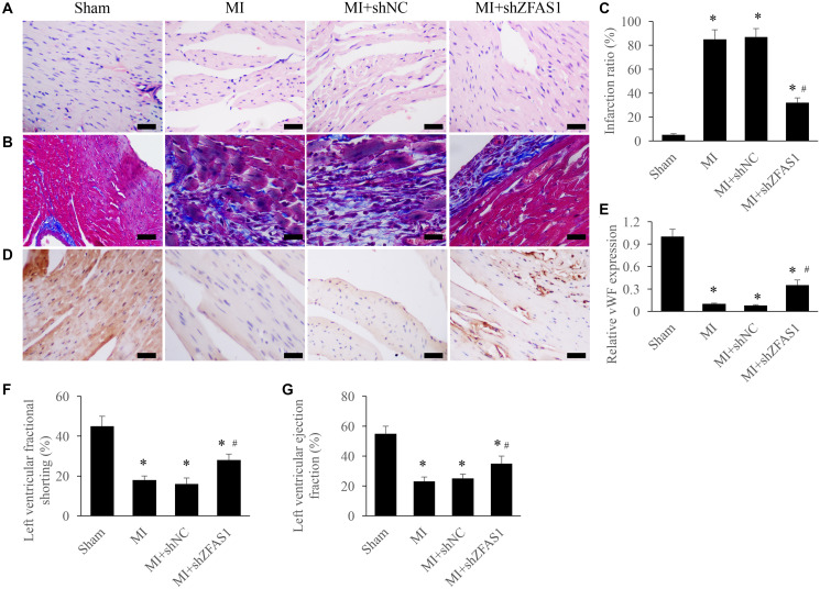
Improvement of cardiac function by silencing ZFAS1 in the MI rats. (A) Influence of shZFAS1 on the histological changes of MI rats myocardial tissues (Scale bar = 500 μm). (B) Influence of shZFAS1 on the collagen deposition of MI rats myocardial tissues (Scale bar = 500 μm). (C) Influence of shZFAS1 on the infarction ratio of MI rats myocardial tissues. (D) The expression of vWF in the MI rats myocardial tissues was measured using IHC staining (Scale bar = 500 μm). (E) Influence of shZFAS1 on the vWF expression in the MI rats myocardial tissues. (F) Influence of shZFAS1 on the left ventricular fractional shortening of MI rats. (G) Influence of shZFAS1 on the left ventricular ejection fraction of MI rats. *P < 0.05 compared with the group sham. #P < 0.05 compared with the group MI.

