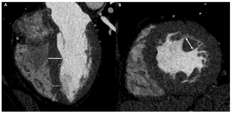
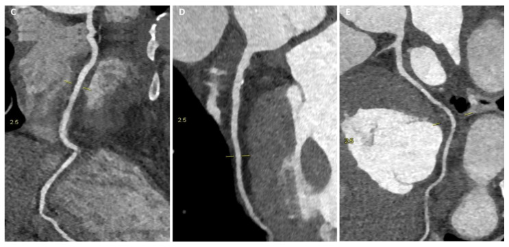
Figure 3.
A 36-year-old male athlete (soccer player) with syncopal episode. CCT images show, in four-chamber plane (A), asymmetric septal hypertrophy with maximum diastolic thickness (16 mm) (full line) and minimum diastolic thickness (11 mm) (dashed line) and, in short-axis plane (B), a hypertrophic papillary muscle (14 mm) (double-headed arrows). With the same scan acquisition, significant stenosis was excluded in the right coronary artery (C), in the anterior descending artery (D), and in the circumflex artery (E).


