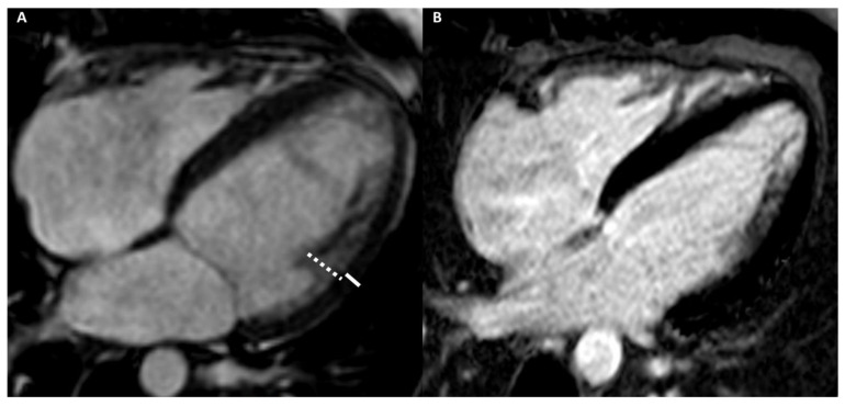
Figure 6.
A 29-year-old male athlete (soccer player) with ECG anomalies. Cine-CMR images show hyper-trabeculation of mid-apical lateral LV wall with the non-compacted wall thickness of 13 mm (dashed line) and the compacted wall thickness of 5 mm (full line) (A). The ratio between non-compacted/compacted was 2.6, with the achievement of the Petersen’s criteria. Delay-MRI image demonstrates the absence of LGE (B). The final diagnosis was LVNC cardiomyopathy.

