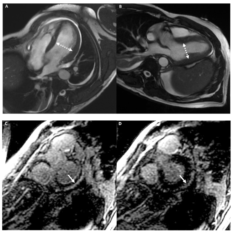
Figure 8.
A 39-year-old male athlete (cyclist) with palpitations. Cine-CMR images show mild LV dilation with increased dimension in four-chambers (A) and in LV outflow-tract (B) plane (dashed double-headed arrows). Delay-MRI contrast images demonstrate, in short-axis planes (C,D), a subepicardial linear LGE at the basal infero-lateral LV wall (arrow). The final diagnosis was LDAC.

