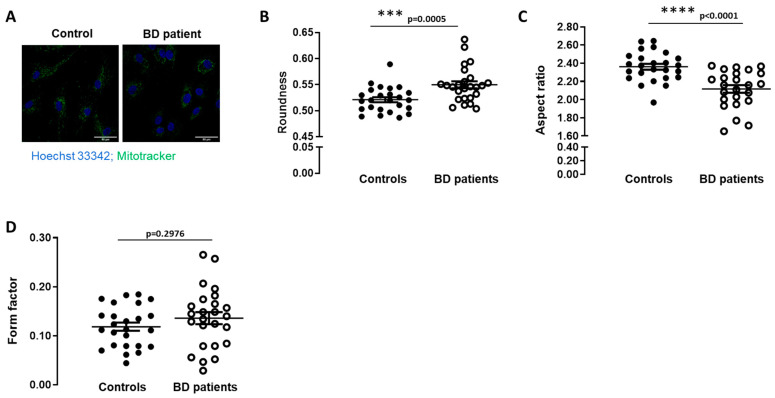
Figure 1.
Mitochondrial morphology in BD patients’ fibroblasts. Mitochondrial morphology was analyzed by fluorescent microscopy using Mitotracker Green. (A) Representative confocal microscopy images of the mitochondrial network shown in green and Hoechst 33342-stained nuclei in blue (magnification, ×40). (B) Mitochondrial roundness, (C) mitochondrial aspect ratio, (D) mitochondrial form factor were determined through the analysis of the fluorescence of confocal microscopy images. Data are the mean ± SEM of 5 different individuals of each group. The experiments were carried out in duplicate and 5 images were analyzed. *** p < 0.001; **** p < 0.0001, significantly different from control group, as determined by Mann–Whitney non-parametric test.

