Figure 4.
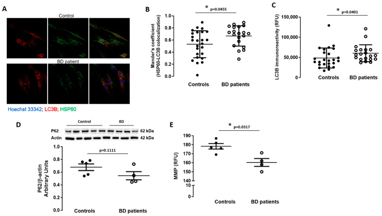
Mitophagy-associated proteins in BD patients’ fibroblasts. Co-localization of LC3B (autophagosome marker) and HSP60 (mitochondrial marker) was evaluated by immunocytochemistry. (A) Representative confocal microscopy images of LC3B (red) and HSP60 (green) immunoreactivity and nuclei labelling with Hoechst 33,342 (blue) (magnification, ×40). LC3B and HSP60 co-localization (B) as well as LC3B staining (C) were quantified using the ImageJ software. The experiments were carried out in duplicate and 5 images were analyzed. The protein levels of p62 (D) were evaluated in fibroblasts from BD patients and control individuals through Western blot analysis. β-actin was used as loading control. The results were normalized to β-actin. The mitochondrial membrane potential (E) was assessed using the fluorescent probe TMRE. Basal fluorescence (excitation: 505 nm; emission: 525 nm) was measured using a microplate reader. Data are expressed as mean ± SEM of 5 different control individuals and 4 BD patients. WB representative images correspond to BD and control samples analyzed on the same gel. * p < 0.05, significantly different from control group, as determined by Mann–Whitney non-parametric test.

