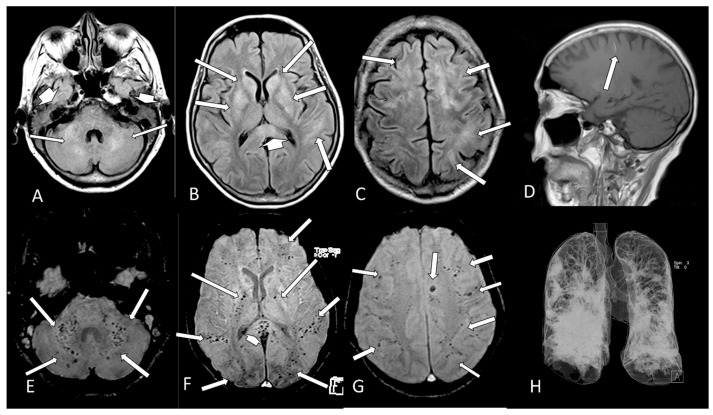
Figure 5.
Case 30—The brain MRI shows multiple and confluent areas of diffuse hyperintensity on FLAIR (A–C, arrows) and liquid in the mastoid cells (A, short head of arrows). A small linear hyperintense subcortical on sagittal T1 (D, arrow), which represents methemoglobin, is observed. There are multiple small dots of micro bleeds at the cerebellum and middle cerebellar peduncle (E, arrows), intern capsule (F, long arrows), splenium of corpus callosum (F, head of arrow) and subcortical white matter (F,G, arrows) of the brain. The 3D chest CT reconstruction shows that there is more than 50% of opacification in both lungs (H).

