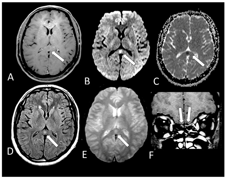
Figure 6.
Case 18—On MRI, there is a small round lesion on the splenium of the corpus callosum which could represent a cytotoxic lesion due to a cytokine storm and differential diagnosis is with small acute infarct. It is hypointense on T1 (A, arrow) with restricted diffusion, being hyperintense on DWI (B, arrow) and hypointense on ADC-Map (C, arrow). This lesion is also hyperintense on FLAIR (D, arrow) and T2* (E, arrow) without microbleeding. The olfactory bulbs are hyperintense on coronal post contrast fat suppressedT1WI (F, arrows) in comparison to the gray matter of the frontal lobes, which can represent enhancement, but we cannot exclude microbleeding.

