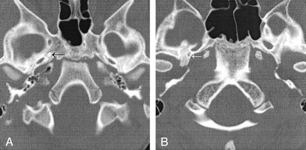
Fig 2.
Absent foramen spinosum.
A, Case 1. CT scan demonstrates bilateral absence of the foramen spinosum. A foramen of Vesalius is noted on this image on the right (arrow) and was present on the left on additional images (not shown).
B, Case 2. CT scan demonstrates a right foramen spinosum on the right (arrow) and absence on the left.

