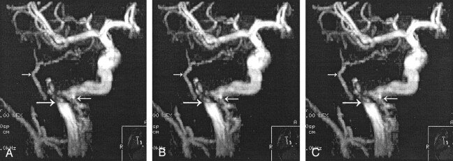
Fig 6.
Case 1. Stereoscopic MRA images of the right carotid. Images A and B are for cross-eyed viewing, and images B and C are for parallel-eyed viewing. The top left arrow shows the PSA supplying the middle meningeal distribution. The right arrow shows the duplicated ICA (carotid branch of the ascending pharyngeal artery). The bottom left arrow points to the turbulent blood flow at the inferior tympanic canal in the aberrant ICA. Views of the left ICA showed similar findings (not shown).

