Fig 1.
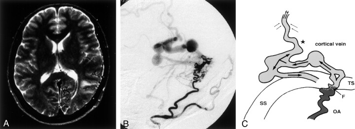
Type 1. Case 7 in a 67-year-old man. with cerebral hemorrhage.
A, T2-weighted MR image reveals no hyperintense lesion in the left temporo-parietal lobe.
B, Left external carotid angiogram, lateral projection, shows DAVFs adjacent to the left transverse sinus. Venous drainage is retrograde into the left transverse sinus. An accessory drainage route into the superior sagittal sinus is recognized. Multiple varices are seen in the venous drainage path.
C, Schematic diagram of a DAVF with an accessory route (star) in the retrograde venous drainage (single arrows). The accessory route with retrograde flow (top double arrows) and the surrounding venous flow (left and right double arrows) drain into another sinus through this accessory route. F indicates the fistula point; OA, occipital artery; SS, sigmoid sinus; and TS, transverse sinus.

