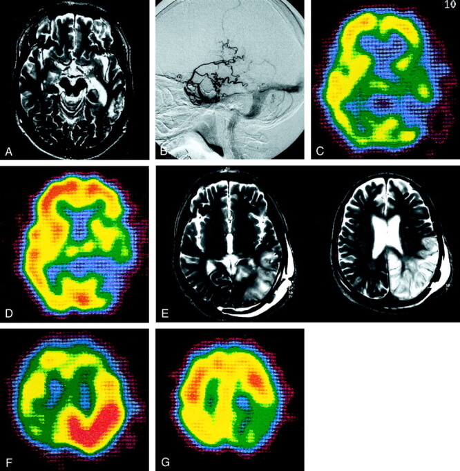
Fig 3.

Type 2b. Case 21 in a 63-year-old man.
A, T2-weighted MR image reveals a hyperintense lesion in the left temporo-occipital lobe.
B, External carotid angiogram, lateral projection, of the left occipital artery shows DAVFs adjacent to the left transverse sinus. No accessory route is recognized.
C, 99mTc-HMPAO SPECT scan shows a hypoperfused area at the site of the lesion.
D, The hypoperfused area is not increased after the acetazolamide challenge.
E, After treatment, the hyperintense area seen on the T2-weighted MR image persists and expands to the left parietal lobe.
F, SPECT image obtained immediately after treatment reveals hyperperfusion in the left parietal lobe.
G, SPECT image obtained 6 months after treatment demonstrates hypoperfusion in the left parietal lobe.
