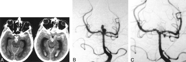
Fig 1.
45-year-old man with sudden severe headache and loss of consciousness was classified as Hunt and Hess grade 5.
A, Axial nonenhanced CT images demonstrate extensive subarachnoid and intraventricular blood.
B, Diagnostic angiogram shows a basilar tip aneurysm. The superior lobulations may represent an associated pseudoaneurysm.
C, Angiogram obtained after coil embolization shows complete occlusion of the aneurysm. There is minimal protrusion of coil loops into the basilar tip, but the patent vessel is widely patent. The patient had a good recovery and at 6-month follow-up was able to resume normal daily life.

