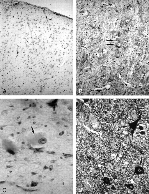
Fig 1.

Photomicrographs show the histologic characteristics of the FCD subtypes.
A, Architectural dysplasia characterized by moderate derangement of cortical lamination, with neurons of the same shape and size scattered throughout the cortex (Kluver-Barrera stain; original magnification, ×250).
B, Cytoarchitectural dysplasia. Note the cluster of cytomegalic neurons with satellitosis (arrows) (Kluver-Barrera stain; original magnification, ×250).
C, Taylor’s FCD with balloon cells. Note large balloon cell characterized by homogeneous eosinophilic cytoplasm and peripheral nucleus with prominent nucleolus (arrow) (hematoxylin-eosin stain; original magnification, ×250).
D, Taylor’s FCD with balloon cells. Note large dysmorphic neurons containing abundant cytoplasmic neurofilaments (arrows) (Bielchowsky stain; original magnification, ×250).
