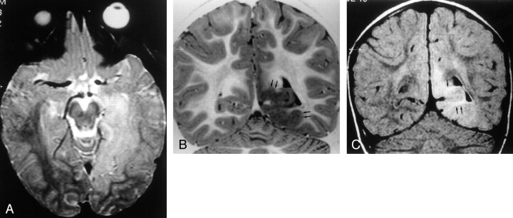
Fig 3.
MR images of Taylor’s FCD without balloon cells.
A, Transverse SE T2-weighted image (2300/90/1) shows extensive hyperintense lesion in the left temporo-occipitobasal region, with no mass effect on adjacent structures.
B, Coronal turbo SE IR T1-weighted image (3000/20/400/2) better demonstrates thickening of the cortex (arrows) with blurring of the gray-white matter junction and subcortical white matter hypointensity.
C, Coronal turbo SE FLAIR T2-weighted image (6000/100/2000/3) reveals that the hyperintensity of the lesion mainly involves the subcortical white matter (arrows). The ventricular trigone is enlarged on the left.

