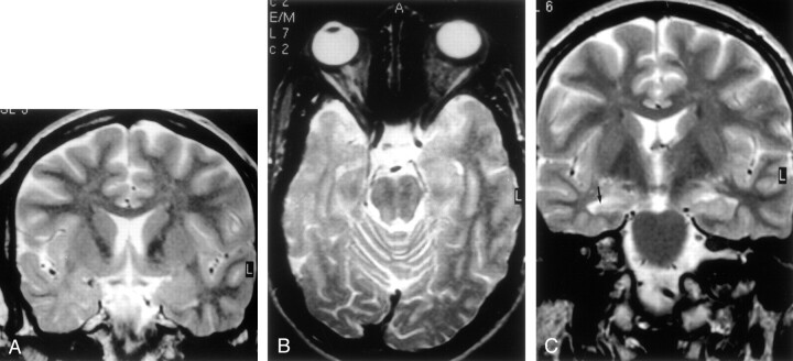
Fig 5.
MR images of architectural dysplasia and ipsilateral hippocampal sclerosis (dual abnormality).
A, Coronal turbo SE T2-weighted image (2300/100/4) reveals hypoplasia of right temporal pole with white matter hyperintensity.
B, Transverse SE T2-weighted image (2300/90/1) confirms reduced volume of right temporal pole compared with the contralateral side, with enlargement of the overlying subarachnoid space.
C, Coronal turbo SE T2-weighted image (2300/100/4) shows right hippocampal head (arrow), characterized by atrophy and signal hyperintensity, suggesting hippocampal sclerosis.

