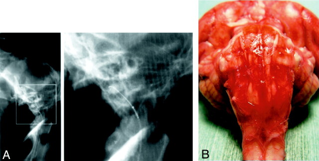
Fig 1.
A, Lateral (left) conventional radiograph of the occipital cranium and cervical spine of a dog demonstrate the microcatheter and microguidewire inserted into the ventral CM. Right part of A is an enlargement of the rectangular area on the left.
B, Photograph of a gross specimen shows the ventral surface of the brain stem after injection of 0.5 ml/kg autologous fresh blood. The BA is completely embedded in clotted subarachnoid blood.

