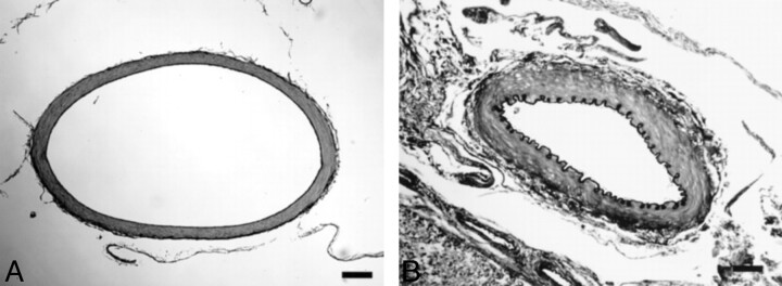
Fig 4.
Light-microscopic appearance of the BA removed at autopsy on the 7th post-SAH day (elastica van Gieson stain; original magnification, x100). Black bars in A and B indicate 100 μm.
A, Physiologic saline (0.5 ml/kg) was injected into the ventral CM. The diameter of the BA is normal.
B, Autologous blood (0.5 ml/kg) was injected into the ventral CM. There is severe narrowing of the vessel diameter, with folding and corrugation of the internal elastic lamina.

