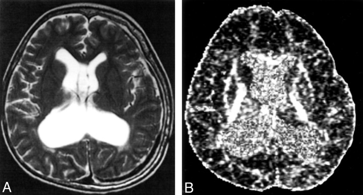
Fig 3.
A 10-year-old medulloblastoma survivor with treatment-induced WM injury and postsurgical complications of hydrocephalus and shunt infection.
A, Axial T2-weighted images (100/4000/2 [TE/TR/NEX]).
B, Axial echo-planar spin-echo DT imaging-derived FA maps (minimum/10000/1200/1 [TE/TR/b factor/NEX]; b = 1200 s/mm2 × 25 directions and b = 0) showing (a) grade 3 white matter changes and (b) reduced signal intensity in the WM compared with the healthy age-matched control, in keeping with reduced FA.

