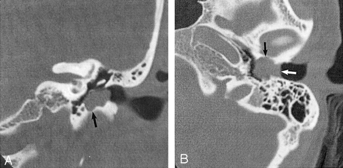
Fig 1.

EACC. Used with permission (24).
A, Coronal temporal bone CT image shows an EACC as a soft-tissue mass in the inferior EAC, with associated erosion of the subjacent bone (arrow). Note the medial bowing of the tympanic membrane in this postsurgical 40-year-old woman with a history of hearing loss.
B, Axial temporal bone CT image in the same patient shows the soft-tissue mass filling the inferior EAC (white arrow), with anterior (black arrow) and posterior EAC erosion. Erosion involving more than one EAC wall is typical.
