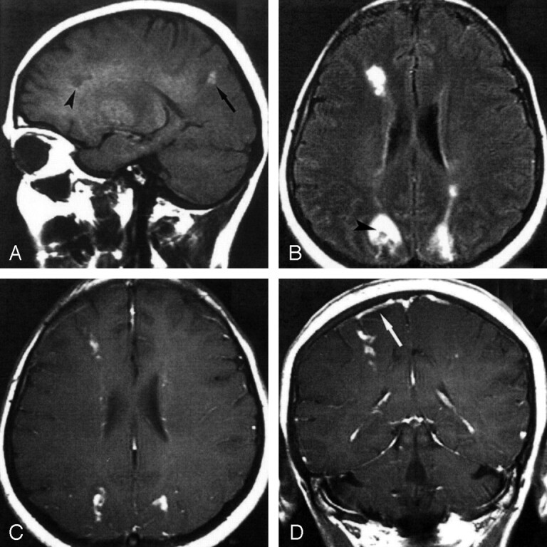
Fig 2.

MR images before treatment.
A, On this nonenhanced sagittal T1-weighted MR image, the occipital lesion appears hyperintense (arrowhead), whereas the frontal lesion is hypointense (arrow).
B, Axial FLAIR MR image shows multiple hyperintense lesions. The center of the right occipital lesion appears hypointense (arrowhead).
C and D, Axial (C) and coronal (D) contrast-enhanced T1-weighted MR images show marked contrast enhancement of the lesions. Note the area of parasagittal meningeal enhancement (arrow) on the right side, close to the frontal-lobe lesion.
