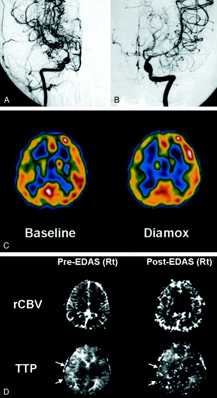
Fig 2.

9-year-old boy with moyamoya disease.
A, Anteroposterior angiogram of the right ICA shows an occlusion of the supraclinoid portion, with development of multiple basal collateral vessels (moyamoya vessels).
B, Left ICA angiogram depicts mild changes of the luminal caliber in the left MCA, but with preserved patency. Left ACA shows severe narrowing.
C, Technetium-99m ethyl cysteinate dimer SPECT scan (left scan) reveals decreased perfusion in both hemispheres, more prominent on the right side. After the acetazolamide (Diamox) injection (right scan), there is no evidence of a significant perfusion reduction.
D, Perfusion MR images show delayed TTP in the right hemisphere before EDAS (arrows in left TTP map) and a signal intensity reduction after EDAS (arrows in right TTP map), which means a restoration of rapid flow at the surgical site. Note, rCBV maps show nothing significant.
