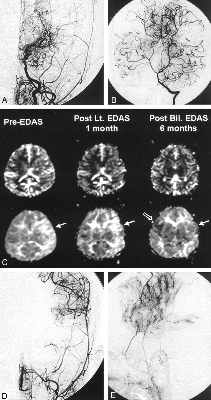
Fig 5.

7-year-old boy with moyamoya disease.
A, Preoperative left carotid angiogram shows an occlusion of the supraclinoid ICA with basal collateral vessels. No transdural collateral vessels from the superficial temporal or middle meningeal arteries are seen.
B, Vertebral artery angiogram shows prominent leptomeningeal collateral vessels supplying the bilateral hemispheres.
C, Serial perfusion-weighted MR images (rCBV, top row; TTP, bottom row) were obtained because the patient underwent bilateral EDAS procedures with a 1-month interval. One-month follow-up perfusion MR image shows a mild reduction in the TTP delay (arrow in middle TTP map), which means neovascularization is beginning to develop. The last perfusion MR image (right TTP map) shows near-complete normalization of the TTP in the left hemisphere (solid arrow) and a partial TTP shortening on the right side (open arrow).
D and E, Postoperative 7-month follow-up left ECA angiograms show successful neovascularization from the middle meningeal and superficial temporal arteries.
