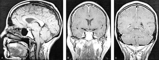
Fig 3.

Contrast-enhanced and unenhanced MR images of the head.
A, Sagittal view T1-weighted image shows “sagging” of the brain with obliteration of the suprasellar cistern, deformity of the interpeduncular cistern and pons, and some descent of the cerebellar tonsils.
B and C, Coronal view contrast-enhanced T1-weighted images show diffuse “felt tip pen” thickening with enhancement.
