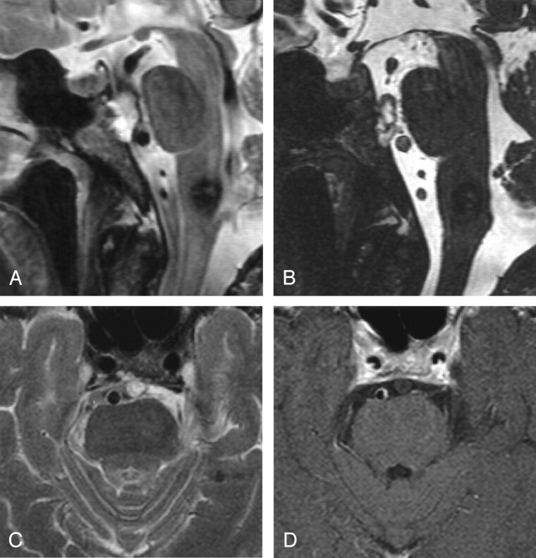
Fig 1.
Patient with known cavernoma in the medulla oblongata.
A, Sagittal T2-weighted image shows an incidental ecchordosis physaliphora at the dorsal wall of the clivus.
B, Intradurally, this is best delineated with a sagittal CISS sequence (3D, 1 mm, TR/TE of 12.06/6.03, flip angle of 70°).
C, Transverse 3-mm T2-weighted image shows hyperintense changes in the dorsal clivus that have a broad connection to the intradural part.
D, No enhancement is seen on this 3-mm contrast-enhanced T1-weighted image.

