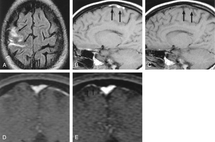
Fig 1.
Patient 1, a 29-year-old woman with headaches, seizures, and cortical venous thrombosis.
A, Axial FLAIR (10,002/158/2200) [TR/TE/TI] MR image shows focal sulcal hyperintensity at the right frontoparietal convexity (arrows).
B, Right parasagittal T1-weighted (500/14) MR image shows tubular hyperintense thrombus (arrows) in a right convexity cortical vein, probably the vein of Trolard.
C, Right parasagittal T1-weighted (500/14) MR image, obtained approximately 3 months after the FLAIR image in panel A, shows resolution of the hyperintense thrombus (arrows).
D and E, Source data from MR venograms obtained at presentation (D) and approximately 3 months later (E) show interval appearance of flow signal intensity (arrows) in the previously occluded cortical vein.

