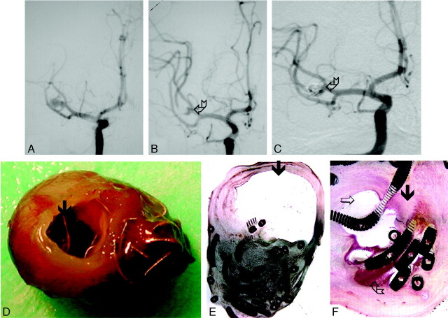
Fig 2.
Ruptured aneurysm of the MCA removed surgically 3 years after treatment with GDCs (case 9, Tables 1 and 2).
A, DSA before treatment.
B, DSA immediately following treatment with standard GDCs, demonstrating neck remnant (broken arrow).
C, DSA 3 years after treatment demonstrates aneurysm recanalization (broken arrow).
D, Gross pathology demonstrating partially exposed coils within the neck (arrow) and coils protruding through the thin wall of the aneurysm dome.
E, Histologic section (H&E stain, low-power magnification, 2×) of the same specimen. Most of the aneurysm sac is filled with organized thrombus, but a large empty space is also seen (arrow).
F, Higher power magnification (20×) demonstrates attenuated fibrocellular tissue (arrow), an empty space (open arrow), and residual unorganized thrombus (broken arrow) within the same aneurysm.

