Fig 3.

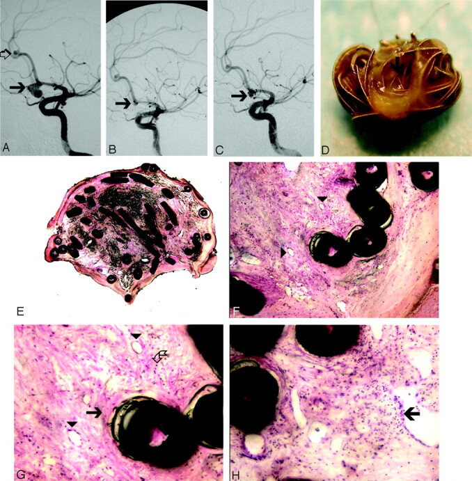
Ruptured aneurysm of the AComA, treated with Matrix coils and removed during surgery 6 months later (case 18, Tables 1 and 2).
A, DSA before treatment demonstrates ruptured AComA aneurysm (arrow) and a small incidental aneurysm at the pericallosal artery (open arrow).
B, DSA immediately after treatment demonstrating a small neck remnant (arrow).
C, DSA, 6 months later, demonstrates growing neck remnant (arrow). Two incidental aneurysms (one on the left MCA and the other on the left pericallosal artery) were clipped, and the AComA aneurysm was clipped and removed during surgery.
D, Gross pathology of the surgical specimen. The coils within the neck (arrow) are covered by a thick tissue layer. The wall of the aneurysm is very thin.
E, Microscopic section of the specimen (H&E stain, low magnification, 2×). The aneurysm cavity is filled with fibrocellular tissue without any residual blood clot or empty spaces.
F, Higher magnification (10×) H&E stain demonstrates coils embedded in fibrocellular granulation tissue with multiple neocapillaries (arrowheads).
G, Higher power view (20×) demonstrates collagen deposition (arrow), smooth muscle cells (broken arrow), and small blood vessels (arrowheads).
H, Leukocyte invasion (arrow) represents granulation tissue (20×, H&E stain).
