Fig 1.

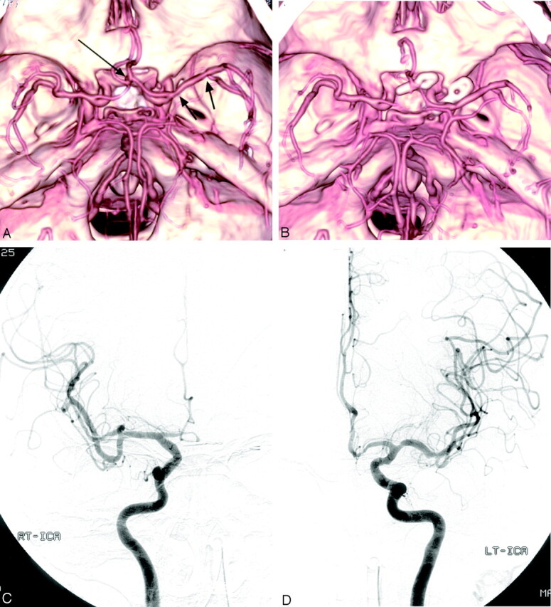
Case 6, a 73-year-old woman with SAH.
A, Preoperative MDCTA image, superior view, shows an anterior communicating artery aneurysm (long arrow). Left posterior communicating artery aneurysm was well visualized on other projections of MDCTA images (not shown). Note the multiple focal stenoses in the left M1 segment (arrows).
B, Postoperative MDCTA image, obtained 7 days after surgery, shows clipping of the aneurysms and multiple spasms of bilateral A1 and A2 segments. Note total occlusion of right A1 segment, as well as no change of stenoses in the left M1 segment consistent with preexisting atherosclerotic stenoses.
C and D, Postoperative right (C) and left (D) carotid angiograms, anteroposterior view, confirm vasospasm involving bilateral anterior cerebral arteries. Note grade 3 spasm (50%–99% luminal narrowing) in the right A1 segment, which was overestimated as grade 4 spasm (total occlusion) on MDCTA (B).
