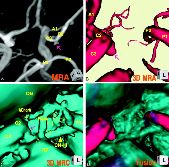
Fig 2.
Case 2, Right infundibular dilation at the junction of the internal carotid artery–posterior communicating artery (fetal-type), in a 36-year-old woman.
A, Maximum intensity projection image of MR angiogram shows a trapezoidal bulging (arrow) at the posterior portion of the supraclinoid internal carotid artery.
B, 3D MR angiogram shows an aneurysm-like protrusion (arrow).
C, 3D MR cisternogram, coordinated projection as to the 3D MR angiogram B, shows an infundibular widening (large arrow) with a large posterior communicating artery (▹), bended at the posterior clinoid process and run tortuously to the posterior cerebral artery. ON, optic nerve; AChorA, anterior choroidal artery (small arrow); PComA (▹).
D, Fusion image of the 3D MR angiography/cisternography shows not an aneurysm but an infundibular dilation.

