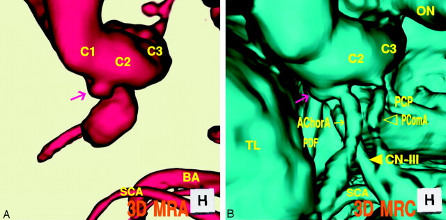
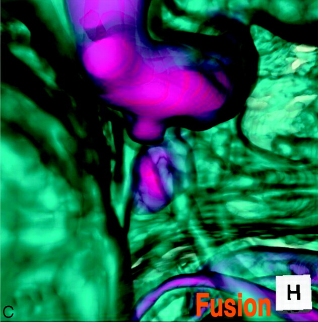
Fig 3.
Case 3, Left infundibular dilation at the junction of the internal carotid artery–anterior choroidal artery, in a 69-year-old-woman.
A, 3D MR angiogram shows an aneurysm-like trapezoid bulging (arrow). C1, the first segment of the internal carotid artery; BA, basilar artery.
B, 3D MR cisternogram, coordinated projection as to the 3D MR angiogram in A, shows an infundibular dilation (large arrow) at the junction of the anterior choroidal artery (small arrow). PComA (▹); CN-III ([GRAPHIC]); TL, temporal lobe.
C, Fusion image of the 3D MR angiography/cisternography shows an infundibular dilation at the junction of an anterior choroidal artery.


