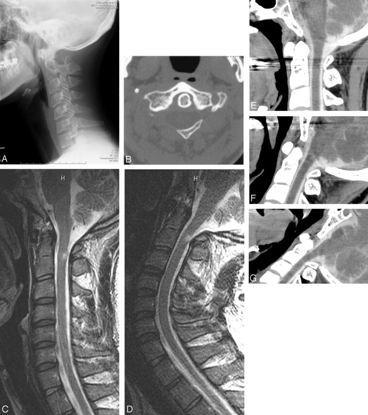
Fig 1.

A, Lateral cervical radiograph shows bilateral bony defects of the posterior arch of the atlas and an isolated posterior tubercle.
B, CT of C1 shows bilateral bony defects of the posterior arch and an isolated posterior tubercle.
C, Sagittal T2-weighted MR image in neutral position shows an intramedullary hyperintensity lesion in the posterior column of the spinal cord slightly below the posterior tubercle of C1.
D, Sagittal T2-weighted MR image in neck extension shows the inward displacement of the posterior tubercle, corresponding to the T2-weighted hyperintensity.
E, MDCT myelography in neutral position shows no abnormality.
F, MDCT myelography in maximal neck extension with the mouth open shows slight ventral displacement of the posterior bone fragment, without cord compression.
G, MDCT myelography during yawning shows cord compression due to the ventral displacement of the posterior bone fragment.
