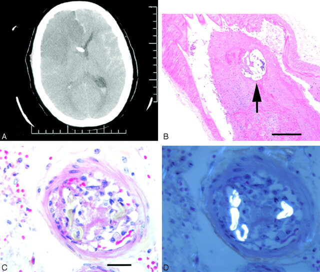
Fig 1.
A, CT scan of patient 1, 2 days after angiography, demonstrating right anterior and middle cerebral artery territory infarcts, as well as a ventricular drain in situ. B, Section of middle cerebral artery containing recent thrombus and a particle of polyvinyl alcohol (arrow, hematoxylin phloxine saffron, ×200). C, Small leptomeningeal artery demonstrating acute cellular reaction and thrombosis and (D) under polarized light, strongly birefringent, hollow fibers characteristic of cotton (C and D, hematoxylin phloxine saffron, ×630).

