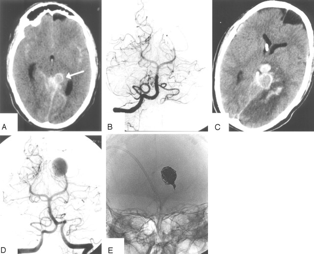
Fig 2.
Patient 20, a 32-year-old man presenting with HH grade III SAH and hemianopsia.
A, CT scan on the day of admission shows SAH and aneurysm in the left ambient cistern (arrow).
B, Vertebral angiogram shows occluded left PCA beyond the P2, presumably by a dissecting aneurysm. Endovascular therapy was judged not necessary.
C, CT scan after sudden clinical detoriation 4 days after admission shows enlargement of the aneurysm, recurrent SAH with thalamic hematoma and hemorrhagic infarction in the PCA territory.
D, Angiogram after recurrent SAH shows filling of large dissecting aneurysm.
E, Occlusion of the aneurysm with coils including the afferent P2. The patient died 3 days later.

