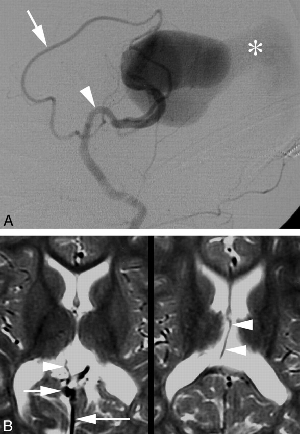
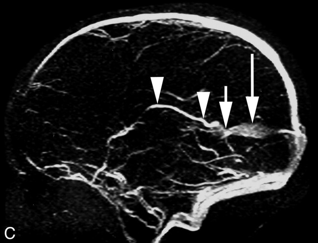
Fig 1.

Five-day-old child with cardiorespiratory failure.
A, Digital subtraction angiography (DSA), left common carotoid artery, lateral view, showing enlarged anterior cerebral (arrow) and anterior choroidal (arrowhead) arteries feeding a vein of Galen aneurysmal malformations (VGAM). Note the drainage of the malformation through a falcine sinus (asterisk).
B, Follow-up magnetic MR imaging 2 years after endovascular therapy; axial T2-weighted images, showing the flow void of a right internal cerebral vein (ICV) (arrowheads) draining into the shrunken vein of Galen (arrow). Note the falcine sinus (long arrow).
C, Two-year follow-up MR venography, sagittal view, showing the course of the right ICV (arrowheads), its termination into the small vein of Galen (arrow), and the falcine sinus (long arrow).

