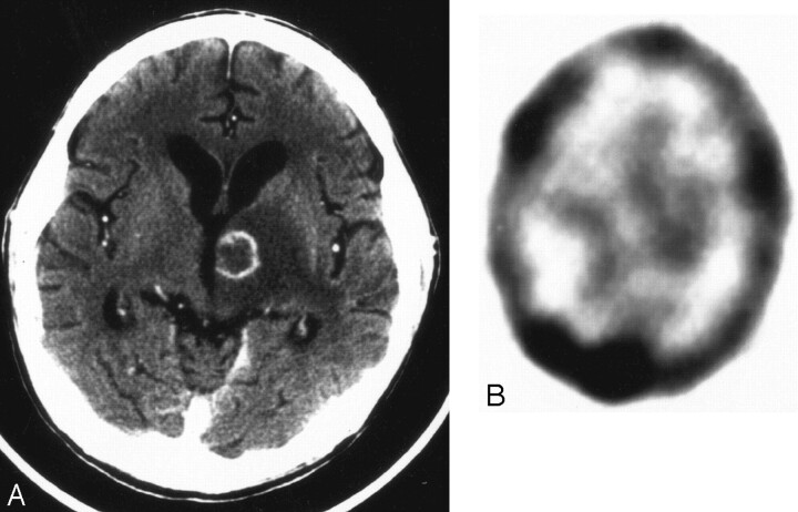
Fig 3.
False-negative thallium-201 SPECT of lymphoma.
A, Axial CT image with contrast shows a 1.9-cm ring-enhancing mass lesion in the left thalamus with surrounding edema.
B, Axial thallium-201 SPECT shows no corresponding area of focally increased tracer uptake. The slight asymmetry in basal ganglia activity (greater on the left) is due to patient angulation (note also asymmetry of the transverse sinus activity, greater on the right). Although suggestive for an infectious etiology, this lesion falls below our size threshold and, by brain biopsy, was due to lymphoma.

