
Fig 2.
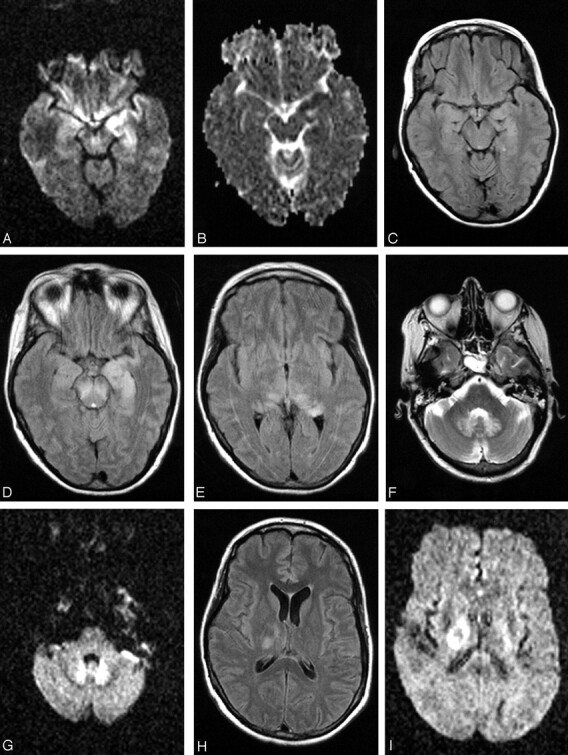
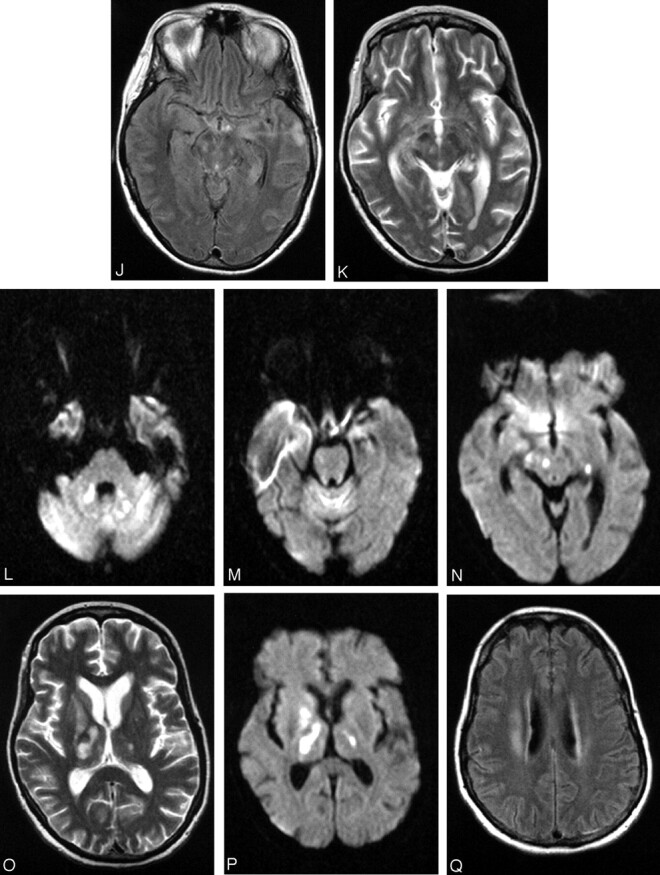
Patient 17, a 49-year-old woman with history of non-Hodgkin lymphoma in remission presented with fever. Work-up was negative for recurrence. The first MR imaging of the brain shows abnormality in the left mesial temporal lobe on trace DW image (A) and ADC (B), though the finding is subtle on the FLAIR sequences (C). At the time of imaging, the findings raised the question of herpes encephalitis, for which the patient was originally treated. Following deterioration of the mental status and development of upper extremity weakness, a second MR imaging was obtained. The abnormality is now apparent on the FLAIR sequences and has progressed to involve not only the contralateral mesial temporal structures, but also the substantia nigra (D), as well as the mesial and dorsal aspect of the thalami (E). Further clinical deterioration with established “polio-like” symptoms prompted a new MR imaging 11 days later, which demonstrated new areas of involvement with resolution of improvement of prior lesions. Images of this MR imaging show increased signal intensity in the dentate nuclei on FLAIR (F) and DW (G) images and right thalamus on FLAIR image (2 hours) and DW image (I) and improvement of the mesial temporal lobe and midbrain abnormalities (J). Focus of T2 hyperintensity is seen in the right red nucleus on FSE T2-weighted image (K). On J, note site of biopsy in the lateral aspect of the left temporal lobe that was negative for herpes encephalitis. The fourth MR imaging was obtained upon patient’s discharge to a nursing facility. DW images show increased signal intensity in the cerebellar hemispheres and right branchium pontis (L), vermis (M), and the right red nucleus (N). Cerebellar hemisphere abnormalities were more conspicuous on DW than FLAIR or FSE T2-weighted images. FSE T2-weighted (O) and DW (P) images through the level of the basal ganglia demonstrate persistent abnormality in the right thalamus and new lesions in the right globus pallidus and left thalamus, whereas the FLAIR image demonstrates involvement of the right corona radiata (Q) as well.