
Fig 3.
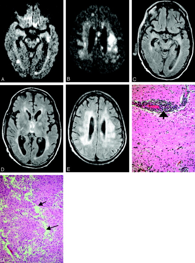
Patient 11, a 51-year-old man post renal and pancreatic transplant with recent mosquito exposure. Axial DW images show patchy areas of increased signal intensity in the periventricular white matter and left cerebral peduncle (A) as well as corona radiata and corpus callosum (B). Axial FLAIR sequences demonstrate subtle abnormalities in the left mesial temporal lobe, both cerebral peduncles, more pronounced in the left one (C), and both thalami and left globus pallidus (D). Diffusely increased signal intensity is also present in the deep hemispheric white matter (D and E). Histopathology (F) shows the hippocampus with marked perivascular (bold arrow) lymphocytic inflammation (hematoxylin and eosin; original magnification 200×). Histopathology (G) demonstrates white matter infarct with necrosis, macrophage and early cavitary changes (cavitation delineated by arrows) (hematoxylin and eosin; original magnification 200×).